Author: Kelly MacKenzie, MD, Emergency Medicine Resident, PGY1
Faculty: Alexis Cates, DO, Medical Toxicology/Emergency Medicine Attending
The Case:
A 3 year old boy with no medical problems presents to the emergency department for cough, fever, and rash. The patient is otherwise healthy, up to date on vaccinations, and was born full-term. Parents report that the patient started complaining of headache and dry cough one week ago and then developed a fever and diffuse rash two days ago. On physical exam, the patient is febrile to 38.5C and a diffuse maculopapular rash with otherwise normal physical exam. Patient is diagnosed with a viral illness and discharged with supportive care. Two weeks later, the patient is brought back to the emergency department with worsening fever, cough, rash and new epistaxis and oral mucosal bleeding. At this time, the patient is noted to be febrile, tachycardic, and hypertensive. The maculopapular rash is consistent with prior visit, but the patient is also noted to have erythema spreading from the distal extremities, starting on the palms and soles of the feet. Labs are drawn due to the patient’s worsening clinical appearance and he is found to have a profound thrombocytopenia with platelets of 4. The patient is admitted and diagnosed with idiopathic thrombocytopenic purpura (ITP, now immune thrombocytopenic purpura). On further conversation with the patient’s parents, you find out that two weeks before symptom onset, the patient had accidentally broken a thermometer containing mercury, which the patient’s mother had vacuumed. Blood and urine mercury levels are drawn and sent out, and Toxicology was consulted.
Learning Point #1: Sources of Mercury Exposure
Mercury is a heavy metal that can cause toxicity in any of its three forms: organic, inorganic, and elemental. The majority of mercury exposures in humans come from elemental mercury, which can be found in a variety of sources including dental amalgams and household thermometers. When ingested, elemental mercury is benign; but when vaporized and inhaled, such as after vacuuming a mercury spill, it is quite toxic. Organic mercury in the form of ethyl mercury and methyl mercury is found in biological sources, primarily fresh and saltwater fish. Inorganic mercury exists as mercury salts, and toxicity is usually due to occupational exposure in industries such as electroplating or chloralkali processing. Inorganic mercury salt exposure historically occurred from cosmetic products where they were used as skin-lightening agents. However, more recently the use of mercury in these products has been banned due to its toxicity.
Learning Point #2: Presentation of Mercury Poisoning
There is a wide range of clinical presentations associated with mercury poisoning depending on the chronicity and type of mercury exposure. Mercury disrupts cellular function by altering protein structure and binding with sulfhydryl and selenohydryl groups, so any cell type can be affected, causing potential impairment of any organ system including renal, central nervous, pulmonary, and endocrine.
Acute exposure to vaporized mercury, such as after vacuuming a significant mercury spill, can cause pneumonitis, even leading to acute respiratory failure in severe cases. Several different rashes have also been described, including a maculopapular rash resembling a viral exanthem and acrodynia, which is an erythematous desquamating rash of the palms and soles that is more specifically associated with mercury poisoning. This has also been referred to as Pink’s disease. In the literature there is also at least one case report of ITP secondary to major exposure of vacuumed mercury spill.

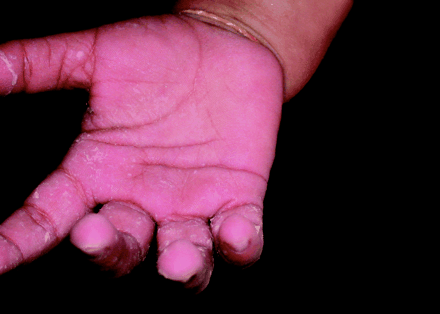
Acrodynia1
Chronic vaporized mercury toxicity frequently presents with non-specific symptoms such as fatigue, gastrointestinal upset and anorexia. Neuropsychiatric manifestations can include erethism, which is a condition characterized by personality changes, excitability or irritability, insomnia, memory loss, and even delirium or hallucinations. Persistent neurological symptoms have been reported even after removal of the exposure. Mercury also inhibits catecholamine-O-methyltransferase (COMT), which decreases the metabolism of catecholamines, causing tachycardia and hypertension which can mimic pheochromocytoma.
The primary manifestation of inorganic mercury poisoning is renal injury which can range from reversible proteinuria to severe acute tubular necrosis. Ingestion of mercury salts is also caustic to the gastrointestinal system and is associated with a variety of symptoms including nausea/vomiting, hypersalivation, and gingivostomatitis.
Organic mercury is particularly concerning in the prenatal period as it is associated with neurodevelopmental and cognitive delays, even causing cerebral palsy in massive ingestion. Exposure outside the prenatal period is associated with neurologic impairment including ataxia, extrapyramidal impairment, and seizures. Severity of symptoms correlates with the amount of exposure.
Case Continued:
The patient’s profound thrombocytopenia is treated with platelet transfusions and steroids with moderate improvement. The mercury blood level returned after several days and was elevated to 161 mcg/L. The family moved out of their home and the Department of Health was notified of the mercury spill. Investigators went to the patient’s home and found evidence of mercury in the carpet at the site of the spill and mercury vapors in the home. The carpet was discarded and the home was properly decontaminated. The patient’s parents and five year old brother were also evaluated for signs and symptoms of mercury toxicity and mercury blood levels were sent. They were completely asymptomatic, but levels returned moderately elevated.
Learning Point #3: Treatment of Mercury Poisoning
The most important step in treating mercury poisoning is removing the exposure. Elemental mercury spills should not be vacuumed under any circumstances as this causes vaporization of the mercury as previously mentioned. Carpets contaminated with mercury must be completely removed and discarded.
Multiple chelation therapies are available for the treatment of mercury toxicity. Thiolated resins including sodium 2,3-dimercaptopropane-1-sulfonate (DMPS), D-penicillamine, and N-acetyl-DL-penicillamine have been used to scavenge mercury in the gastrointestinal tract and have been shown in studies to significantly decrease the blood half-life of mercury. DMPS is the preferred agent used outside the United States while dimercaptosuccinic acid (DMSA) is more commonly used in the United States. Studies evaluating chelating agents have studied DMPS and the results have been extrapolated to DMSA. There is limited information available on the effectiveness and the possible adverse effects of chelation therapy. Some physicians have used elevated mercury blood or urine levels to guide treatment. However, these levels can vary significantly depending on the type, chronicity, and mechanism of mercury exposure and may not accurately represent total body burden. Without a clear history of known mercury exposure, it can be difficult to know how severe the exposure was. Therefore, improvement in mercury levels may not be clinically useful in determining if chelation therapy is indicated. Furthermore, while some short term studies have shown no adverse outcomes with the use of chelation therapy, long term effects of chelation therapy in the context of mercury poisoning have not been fully evaluated. A reasonable approach would be to consider chelation therapy when there has been a known significant mercury exposure causing critical illness. Further research is required to better develop treatment guidelines for chelation therapy in the context of mercury poisoning.
Case Resolution:
Due to the critical thrombocytopenia and known mercury exposure, chelation therapy with DMSA is considered for this patient. DMSA is held until the platelet count increased to 21 after transfusion and immunoglobulin treatment, after which the patient is treated with five days of DMSA. Patient’s symptoms improved significantly with treatment and he was discharged home after 13 days of hospitalization. The patient’s family was not treated with chelation therapy as they remained asymptomatic. Mercury blood levels were retested after removal of the exposure and returned to normal levels.
Resources:
- Weinstein, Michael, and Stacey Bernstein. “Pink ladies: mercury poisoning in twin girls.” Canadian Medical Association Journal, vol. 168, no. 2, 2003, p. 201. https://www.cmaj.ca/content/168/2/201.long.
- Schwartz, Joyce, et al. “Toxicity of a Family From Vacuumed Mercury.” American Journal of Emergency Medicine, vol. 10, no. 3, 1992, pp. 258-261.
- Bernhoft, Robin. “Mercury Toxicity and Treatment: A Review of the Literature.” Journal of Environmental & Public Health, vol. 2012, 2012, pp. 1-10.
- Mckay, Charles. “Public Health Department Response to Mercury Poisoning: The Importance of Biomarkers and Risks and Benefits Analysis for Chelation Therapy.” Journal of Medical Toxicology, vol. 9, no. 4, 2013, pp. 308-312.
- Chan, Thomas. “Inorganic mercury poisoning associated with skin-lightening cosmetic products.” Clinical Toxicology, vol. 49, 2011, pp. 886-891.
- Torres, Alfonso, et al. “Mercury Intoxication and Arterial Hypertension: Report of Two Patients and Review of the Literature.” Pediatrics, vol. 105, no. 3, 2000, p. e34.
